Adaptive Hybrid Optimizer based Framework for Lumpy Skin Disease Identification
Ubaid Ullah*, Department of Computer Science, NUST Balochistan Campus
Quetta, Pakistan, obaidullah@nbc.nust.edu.pk
Muhammad Abid Hussain*, Department of Computer Science, NUST Balochistan Campus
Quetta, Pakistan, engr.abid@nbc.nust.edu.pk
Moid Sandhu*, Australian e-Health Research Centre (AEHRC), CSIRO
Brisbane, Australia, moid.sandhu@csiro.au
Abd Ullah Khan*, CQI Lab, Department of Electronics and Information Convergence Engineering, Kyung Hee University, Global Campus
Gyeonggi-do, Korea (south), abdullah@khu.ac.kr
Muhammad Azfar Yaqub*, Faculty of Engineering, Free University of Bozen-Bolzano
Bolzano, Italy, myaqub@unibz.it
Mohsin Raza Jafri*, Department of Computer Science, NUST Balochistan Campus
Quetta, Pakistan, mjafari@nbc.nust.edu.pk
* Corresponding author
Abstract — Lumpy Skin Disease (LSD) is a contagious viral infection that significantly deteriorates livestock health, thereby posing a serious threat to the global economy and food security. Owing to its rapid spread characteristics, early and precise identification is crucial to prevent outbreaks and ensure timely intervention. In this paper, we propose a hybrid deep learningbased approach called LUMPNet for the early detection of LSD. LUMPNet utilizes image data to detect and classify skin nodules– the primary indicator of LSD. To this end, LUMPNet uses YOLOv11, EfficientNet-based CNN classifier selected for its native accuracy–efficiency trade-off, and a novel adaptive hybrid optimizer. More precisely, LUMPNet detects and localizes LSD skin nodules and lesions on cattle images. It exploits EfficientNet to classify the localized cattle images into LSDaffected or healthy categories. To stabilize and accelerate training, we apply the proposed AWDR optimizer independently when training each of the two sequential components of the pipeline: it is used to train YOLOv11 for nodule detection, and separately to train the EfficientNet classifier for LSD-versushealthy classification. The two networks are trained separately, with the output of the detection stage feeding the classification stage only at inference time. We evaluate LUMPNet at various stages of LSD using a publicly available dataset. On a held-out validation set, LUMPNet achieves 98% classification accuracy with 0.99 precision, recall, and F1-score, outperforming the closest directly comparable prior image-based LSD detection method (MobileNetV2 with RMSProp, 95% accuracy on 793 images) by 3% on a similarly sized dataset (1,024 images). The results show that LUMPNet achieves superior performance.
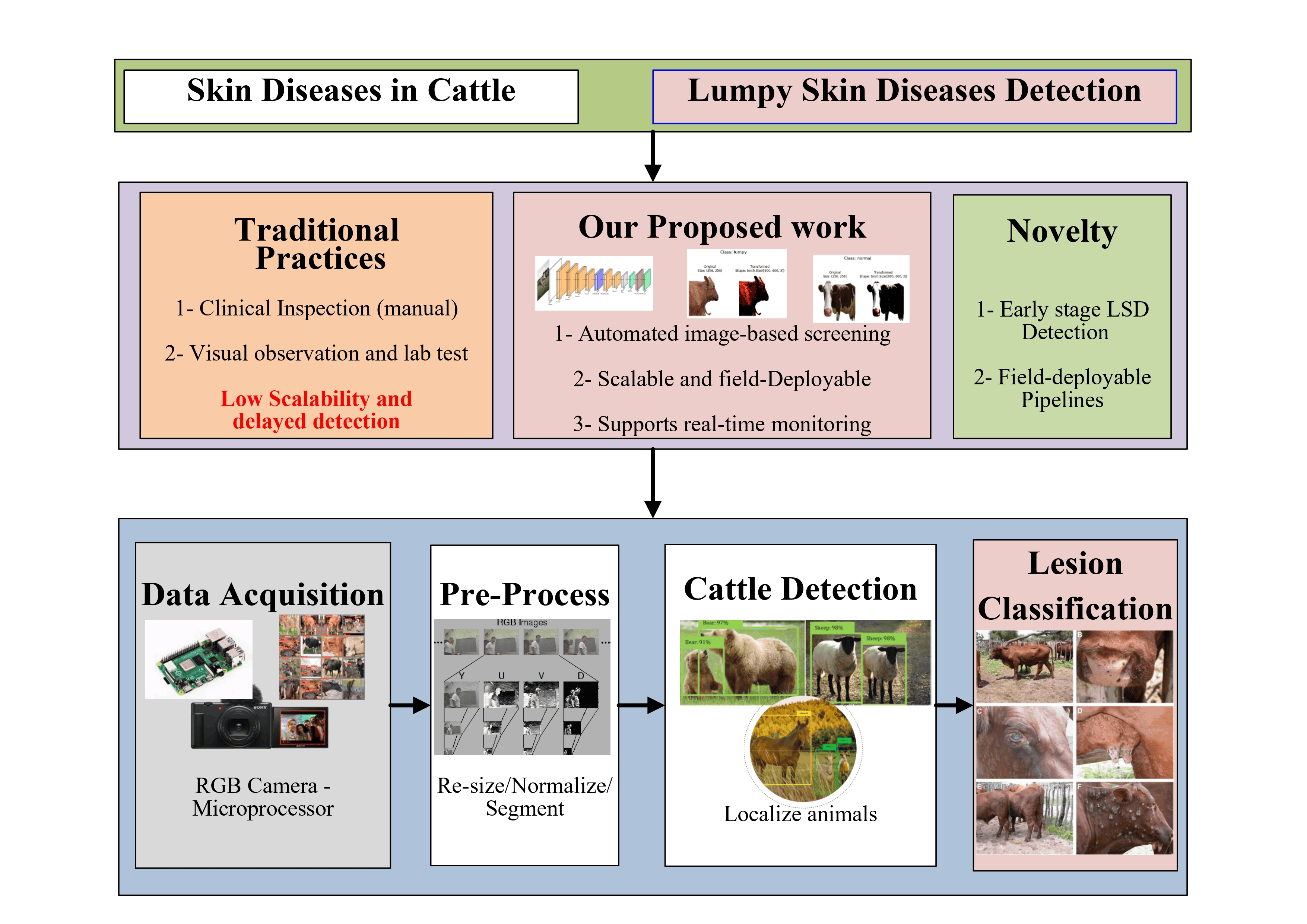
Precision livestock farming utilizes automated structures to monitor livestock, enhancing their production, reproduction, health, welfare, and environmental impact [1]. It plays a vital role in the early detection of diseases and contamination in animals. Specifically, pores and skin parasitic infections pose a chief financial undertaking within the livestock enterprise. These infections cause discomfort, impairing the animal’s capacity to relax and eat well. Moreover, those infections also compromise the skin’s innate defense mechanisms, which in turn increase susceptibility to bacterial infections. Certain ectoparasites, which include blood-feeding flies and precise tick species, are economically significant as they transmit diverse illnesses. The pores and skin lesions attributable to those parasites are specifically harmful. Livestock, consisting of farm animals, cows, and buffalo, are vulnerable to a viral infection referred to as Lumpy Skin Disease (LSD) [2]. This disease is transmitted by means of blood-feeding insects like ticks, flies, and mosquitoes. In addition to causing fever and skin nodules, the virus can be fatal, especially for animals that have never been exposed to it before. Control measures include vaccination and culling infected animals. LSD results in considerable financial losses, affecting species such as cows and water buffalo. The disease is caused by the LSD virus, which belongs to the Capripox genus within the Poxiviridae family. There are several cattle diseases associated with skin nodules, which can be classified based on key characteristics such as the description of nodules, fatality rate, early detection indicators, and the importance of nodule detection. LSD stands out because of its high fatality rate and the extreme nature of the skin nodules, which can be commonly observed on the neck and lower back. Early detection of LSD is crucial to save from its rapid unfold, with signs such as fever, swelling, and reduced milk yield serving as key indicators. Adamu et al. [3] conducted an experimental analysis of LSD-affected cattle in Ethiopia. They confirmed LSDV as the primary cause of the outbreak and identified nodules and fever as the main symptoms of the disease. Recent advancements in artificial intelligence (AI) have also made it possible to perform the early detection of various livestock diseases. In this direction, various researchers have reported their work in the literature related to precision livestock farming. Gouda et al. [4] predicted LSD using multiple ML models under multiclass balance. They apply undersampling and oversampling to address the class imbalance. Random forest along with random oversampling achieved 82% accuracy. LSD is characterized by fever, swollen superficial lymph nodes, and numerous nodules ranging from 2 to 5 centimeters in diameter on the skin and mucosal membranes [9]. Infected cattle also suffer from lameness and swelling in their limbs. The virus often causes lasting skin damage in affected animals. The disease leads to poor growth, infertility, abortion, chronic weakness, decreased milk production, and even death. Fever typically appears about a week after the viral infection, with the initial fever possibly exceeding 41°C and lasting for up to a week. During this period, all superficial lymph nodes become enlarged. The distinctive nodules appear 7 to 19 days after infection, accompanied by mucopurulent discharge from the nose and eyes. These nodules affect deeper layers of skin, such as the subcutis and even the muscle, impacting both the dermis and epidermis. The lesions may be well-defined or merge together and can appear anywhere on the body, though they are most commonly found on the head, neck, udder, scrotum, vulva, and perineum. Skin lesions may either resolve quickly or persist as painful lumps. In some cases, the lesions may become more severe, forming deep ulcers that may be pusfilled and surrounded by granulation tissue. A growing body of research is leveraging ML to predict the occurrence of infectious diseases in animal populations. Ananda et al. [10] proposes using deep CNN feature extractors i.e. VGG-16, VGG-19, Inception-v3, combined with machine-learning classifiers for automated LSD detection, reporting improved feature extraction and better performance versus classical methods. Raj et al. [11] proposed a hybrid model based on VGG19 and ResNet-50 deep learning architectures to accurately detect LSD conditions. They used principal component analysis to decrease the dimensionality of the feature vector. Liang et al. [12] utilized machine learning techniques to predict outbreaks of African Swine Fever by analyzing global bioclimatic data. While other methods achieved an accuracy of 80.4%, the Random Forest technique proved more accurate than the SVM when applied to a dataset of key climate variables, with an accuracy of 76.02%. Figure. 1 summarizes the role of our proposed LUMPNet in Precision livestock farming and early disease detection.
Figure 1. Summary of proposed work
Table 1. Comparison of our work with rekated works
Ref. (Year)
Approach
Model Performance
Dataset
Evaluation Methods
Challenges
Girma et al. (2021)
CNN for feature extraction and SVM for classification in LSD detection.
95.7% accuracy with SVM for LSD classification.
1,740 images for LSD detection.
Evaluated using SVM and Random Forest, with SVM achieving 95.7% accuracy.
Noise removal techniques are needed to improve image quality and region identification.
Ujjwal et al. (2022)
Random Forest for LSD prediction in specific geographic locations.
97% accuracy with Random Forest.
18,603 instances and 16 features.
Performance comparison using Random Forest, achieving 97% accuracy.
Image noise and challenges in accurate segmentation for LSD detection.
Senthilkumar et al. (2024)
Automated LSD detection using deep learning and transfer learning.
VGG16 and MobileNetV2 achieved accuracies of 96.07% and 96.39%, respectively.
Publicly available datasets of healthy and LSD-affected cattle.
Evaluation using accuracy, sensitivity, specificity, and precision.
Dataset imbalance and the need for high sensitivity.
Saqib et al. (2024)
Deep learning using MobileNetV2 and the RMSprop optimizer.
95% accuracy for LSD detection.
464 healthy and 329 LSD-affected cattle images.
Evaluation of classification performance on healthy and LSD-affected cattle images.
Balanced dataset partitioning and image diversity.
Ullah et al. (2025)
Vision Transformer (ViT) classifier with resizing, normalization, and data augmentation.
98% accuracy.
8,000 cattle images.
Evaluation using accuracy, precision, recall, and F1-score.
Ensuring generalization to real-world field conditions.
Proposed: LUMPNet (2026)
YOLOv11 + EfficientNet + AWDR optimizer for early LSD detection and classification.
98% accuracy, with 0.99 precision, recall, and F1-score.
1,024 images.
Evaluation using accuracy, precision, recall, and F1-score on a held-out evaluation set.
Handling early-stage lesion variability and ensuring precise lesion localization.
A. Novelty and contributions
A number of deep learning (DL)-based models have been proposed in the literature for early detection of LSDaffected cattle [7]. However, most of these models do not generalize across diverse environments and data scarcity, leading to suboptimal performance. Additionally, the traditional feature extraction methods adopted by the recent literature fail to effectively classify LSD cases, particularly in the early stages when symptoms, such as skin nodules, are less pronounced. Moreover, most of these models take a long converge time and are not scalable, making them inefficient to be deployed in real-world livestock monitoring systems [13]. To address these issues, we propose a unified, fast-converging, and scalable deep-learning pipeline approach called LUMPNet, which integrates advanced object detection, classification, and intelligent optimization techniques to enhance the accuracy of early-stage LSD detection by focusing on the visual appearance of skin nodules. The modular design of LUMPNet enables it to support multidomain adaptability, enabling re-training for new object categories or environmental conditions with minimal overhead. The novelty and cotributions are as follows. LUMPNet combines YOLOv11-based detection with an EfficientNet-based CNN classifier for accurate localization and precise classification of the affected animals. An Adaptive Hybrid optimizer (AWDR) is proposed, which combines the variance smoothing capability with the decoupled weight-decay regularization of AdamW through a dynamic blending coefficient. The optimizer enhances gradient stability, accelerates convergence, mitigates overfitting, and improves generalization on small or imbalanced datasets. Table 1 summarizes the comparison of LUMPNet with the existing approaches. The rest of the paper is structured as follows. Section 2 presents the proposed model and methodology. Section 3 explains the results, and Section 5 concludes the paper.
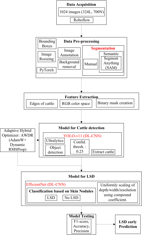
Figure 2. Proposed system architecture
Table 2. Extracted features for LSD detection
Feature Name
Description
Type
Nodule Count
Total number of visible nodules detected on the skin surface.
Quantitative
Nodule Size
Average area or diameter of individual nodules.
Quantitative
Nodule Shape
Circularity, aspect ratio, and irregularity of nodules.
Morphological
Edge Density
Density of edges (sharp intensity transitions) around nodules.
Structural
YOLOv11 Region Proposals
Bounding-box features, including position, size, and confidence score.
Learned Feature
EfficientNet Embeddings
Deep-learned features extracted from intermediate convolutional layers.
Learned Feature
II. Methodology
LUMPNet detects LSD in cattle by integrating deep learning models i.e. YOLOv11 and EfficientNet, with novel AWDR optimizer. More precisely, the model uses a multifaceted approach encompassing object detection and semantic segmentation techniques. The process begins by identifying the cattle in the dataset [14]. Both manual and automated segmentation is performed using the segment anything (SAM) package [15]. This results in separating the cattle’s image from the colored background. Next, the main phase starts, where the symptoms of LSD, specifically skin nodules and lesions, are identified using YOLOv11 [16]. The LSD-affected cattle is identified by finding the skin nodules or the affected skin. For classification, the model utilizes EfficientNet [17]. Figure 2 illustrates the system architecture, which comprises the following stages: dataset preparation, training a YOLOv11 model, segmentation, training an LUMPNet model for LSD classification, and evaluation. In this paper, we use image dataset publicly available on Kaggle [14]. The dataset contains 1024 cattle images, in which both healthy and LSD affected cattle images exist. In these images, the LSD affected cattle have a mild to extreme appearance of skin nodules. The appearance of nodules characterizes the multiple stages of LSD. Among these images, there are 324 images of cattle with lumpy skin and an additional 700 images of healthy cattle. A key challenge was to develop an effective partitioning strategy that would support accurate model training and evaluation. To this end, 500 images of healthy cattle were allocated for training, while the remaining 200 were designated for testing. Similarly, 224 images of LSD-affected cattle were selected for training, and 100 images were reserved for testing.
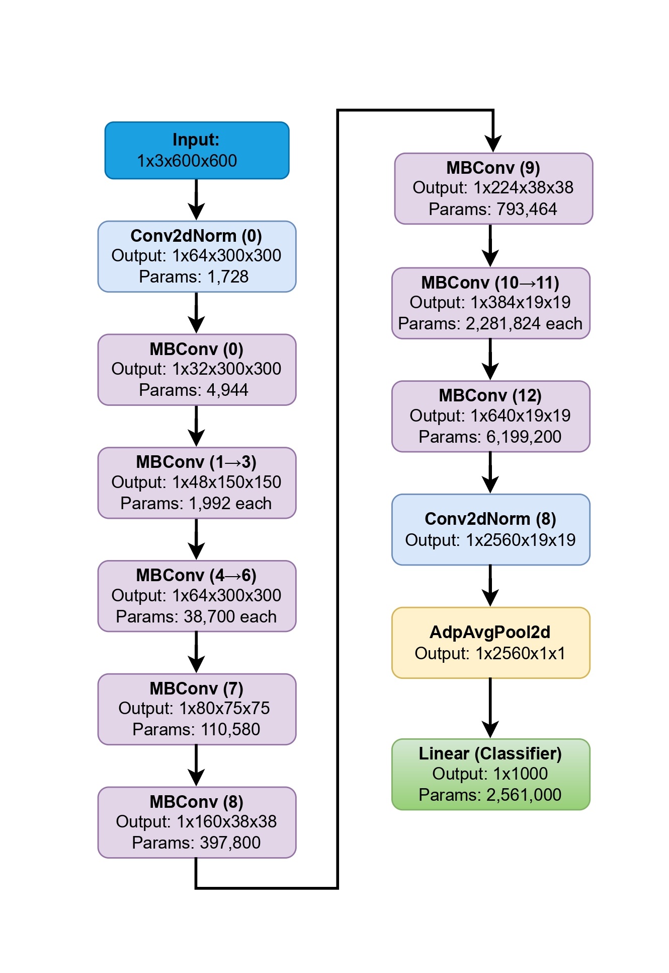
Figure 3. EfficientNet Model Layer Dimensions
A. Image Pre-processing
During image pre-processing, we perform both manual and automatic segmentation. Moreover, we mask the background color of the images with blue to maintain consistency, enhance the focus of the cattle’s skin, and ultimately improve the accuracy of the model. This leads to the accurate detection of skin-related features. For automatic segmentation, we utilize SAM, which provides more accurate region detection. We improve model prediction by visualizing masks, points and bounding boxes through the utility functions.
B. Feature Extraction
The feature extraction stage operates on the cattle region obtained after pre-processing/segmentation and produces three complementary cues that are fed to the learning models as explained in Table 2. Specifically, we compute (1) Edges of cattle: gradient-based edge maps to emphasize nodule boundaries and local shape irregularities; (2) RGB color space: the native RGB representation with per-channel statistics that preserve appearance and pigmentation differences associated with lesions; and (3) Binary mask creation: a foreground mask isolating the cattle silhouette to suppress background clutter and constrain subsequent analysis. Formally, for an input image x and its segmentation ˆ M, we derive E = edge(x⊙ ˆ M), C =xRGB, and B =⊮( ˆM >0), and form a composite feature tensor F = [E, C, B]. During training, F is paired with image-level labels (healthy vs. LSD-affected) to supervise the models; at test time, the same features guide prediction. The above-mentioned features provide complementary geometric and photometric evidence that improves nodule localization.
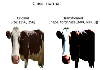
Figure 4. Images showing the accuracy of EfficientNet in transforming Class: Normal cattle
C. Implementation of YOLOv11
YOLOv11 is a CNN-based framework for real-time object classification and detection [16]. It introduces architectural enhancements that include Cross Stage Partial with kernel size 2 (C3k2) blocks for efficient feature reuse and Spatial Pyramid Pooling-Fast (SPPF) for multi-scale context. It strengthens spatial focus using Convolutional block with Parallel Spatial Attention (C2PSA). These characteristics enhance performance, particularly for detecting subtle LSD skin nodules in cattle. We implement pretrained weights via transfer learning and generate detection plots for qualitative evaluation. YOLOv11 partitions an input image into multiple grid cells. Each cell predicts bounding boxes with associated confidence scores. During bounding box regression, the regression model predicts these parameters via the Distribution Focal Loss (DFL) mechanism, permitting localization of LSD lesions on livestock pores and skin. Residual connections permit the community to study complicated capabilities vital for subtle lesion detection. YOLOv11 utilizes Complete IoU (CIoU) loss that promises high-precision lesion localization. DFL also captures uncertainty in bounding box localization, particularly beneficial for detecting LSD lesions with ambiguous or blurry boundaries. Binary Cross-Entropy (BCE) loss optimizes class predictions for each detected object. The attention mechanism allows YOLOv11 to concentrate computational resources on potentially lesioncontaining regions while suppressing irrelevant background features, dramatically improving detection accuracy for small or partially occluded LSD nodules, a common challenge in field conditions where cattle have varying coat colors, lighting conditions, and occlusions from environmental factors. The model architecture continues to grow deeper with more convolutional blocks and feature fusion operations through the neck component, enabling the extraction of both low-level and high-level features. Lower-level features extracted in earlier layers focus on fine details, including edges, texture patterns, color variations, and subtle skin surface changes characteristic of early-stage LSD lesions.
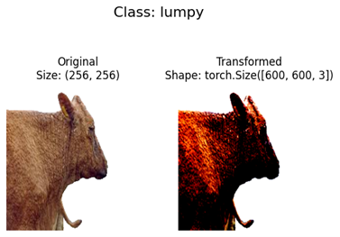
Figure 5. Images showing the accuracy of EfficientNet in transforming Class: LSD affected cattle
D. Adaptive Hybrid Optimizer: AWDR
We devise an AWDR optimizer that combines the weight decay regularization capabilities of AdamW with the adaptive variance smoothing. The proposed fusion model not only enables stable gradient updates during the early stages of training, moreover, preserves better generalization in later epochs. The optimizer implements a dynamic transition schedule. In this scheduling mechanism, time-dependent coefficient β(t) controls the gradual shift. This optimizer results in more stable convergence and reduced overfitting. It also provides three main advantages over traditional singular optimizer, i.e., Improved training stability across long training cycles.
E. EfficientNet-based Classification
EfficientNet is used as the main component for LSD detection in our model as demonstrated in Figure. 3. It is a convolutional neural network (CNN) architecture known for achieving an effective balance between classification accuracy and computational efficiency. A key built-in feature of EfficientNet is compound scaling, in which the network width, depth, and input resolution are scaled in a unified and balanced manner. The novelty of LumpNet lies in the integration of YOLOv11 for detection, EfficientNet for classification, and the proposed adaptive hybrid optimizer. To stabilize and accelerate training, we apply the proposed AWDR optimizer independently when training each of the two sequential components of the pipeline: it is used to train YOLOv11 for nodule detection, and separately to train the EfficientNet classifier for LSD-versus-healthy classification. The two networks are trained separately, with the output of the detection stage feeding the classification stage only at inference time. This model approaches anomalies in cattle pictures with the use of transfer learning, which allows the model to quickly adapt to the primary features of the dataset. Therefore, it serves as a pretrained version for anomaly detection. Moving in advance with anomaly detection of the use of EfficientNet, the proposed model involves a couple of key steps. These consist of pre-processing responsibilities, which comprise of resizing images to the preferred input proportions for the model and augmenting the dataset to develop model generalization. We alter the model’s parameters on the training dataset to adapt it for LSD detection. To compare the model, we make predictions on the test dataset. The output layer of the model generates the classification results, which can be adjusted to handle specific tasks like distinguishing between LSD-affected cattle and healthy cattle.
Figure 6. LUMPNet Model Accuracy
Table 3. Comparison with similar studies
Model
Accuracy
Extreme Learning Machine (ELM)
92%
DenseNet121
90%
CNN Model
89%
MobileNetV2
84%
RMSProp + MobileNetV2
95%
Proposed LUMPNet
99%
Table 4. Dataset Summary
Metric
Value
Total Samples
1,250
Training Samples
1,000 (80%)
Testing Samples
250 (20%)
Healthy Samples
625
Infected Samples
625
Class Balance Ratio (Healthy:Infected)
1:1.00
III. Results and Discussion
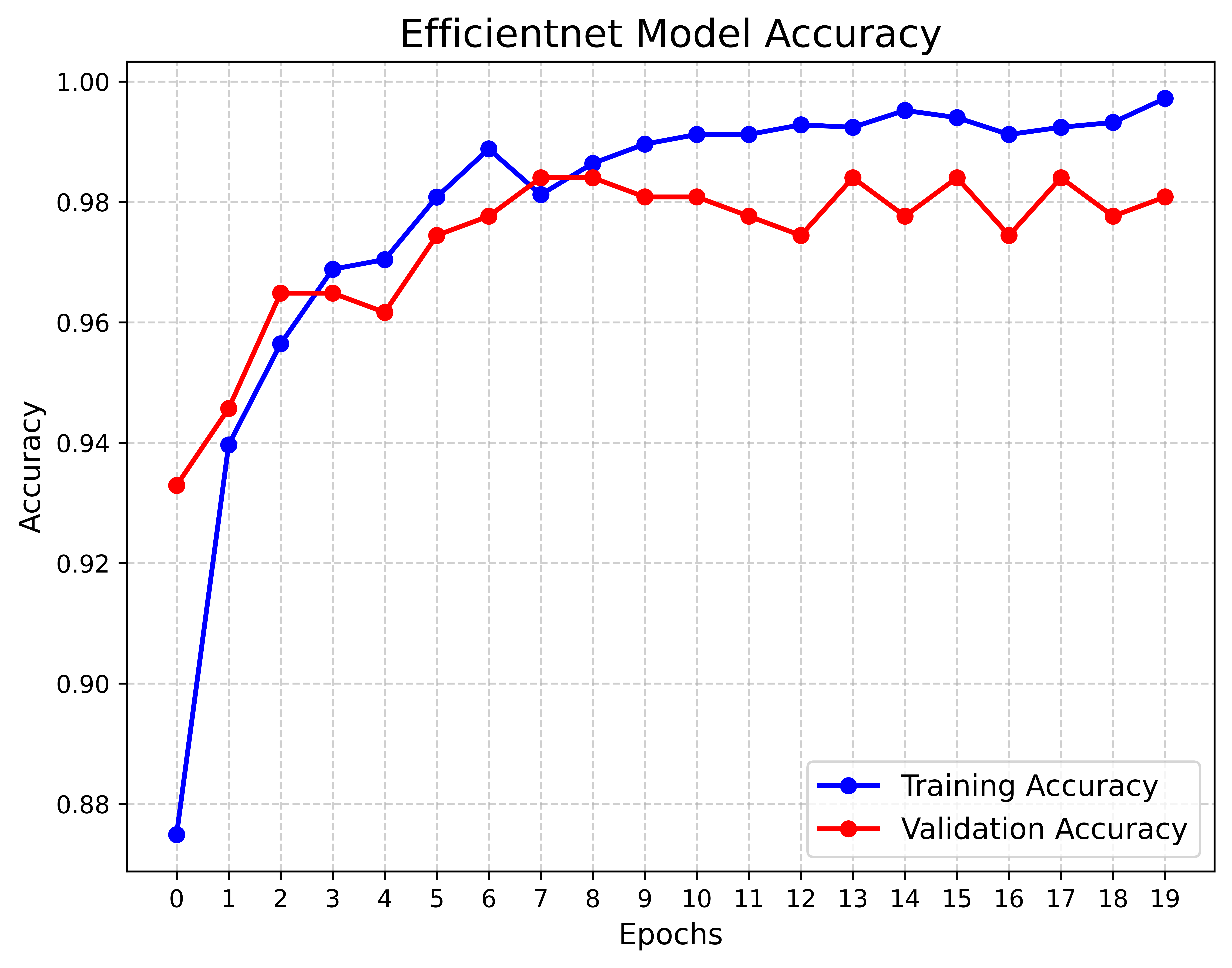
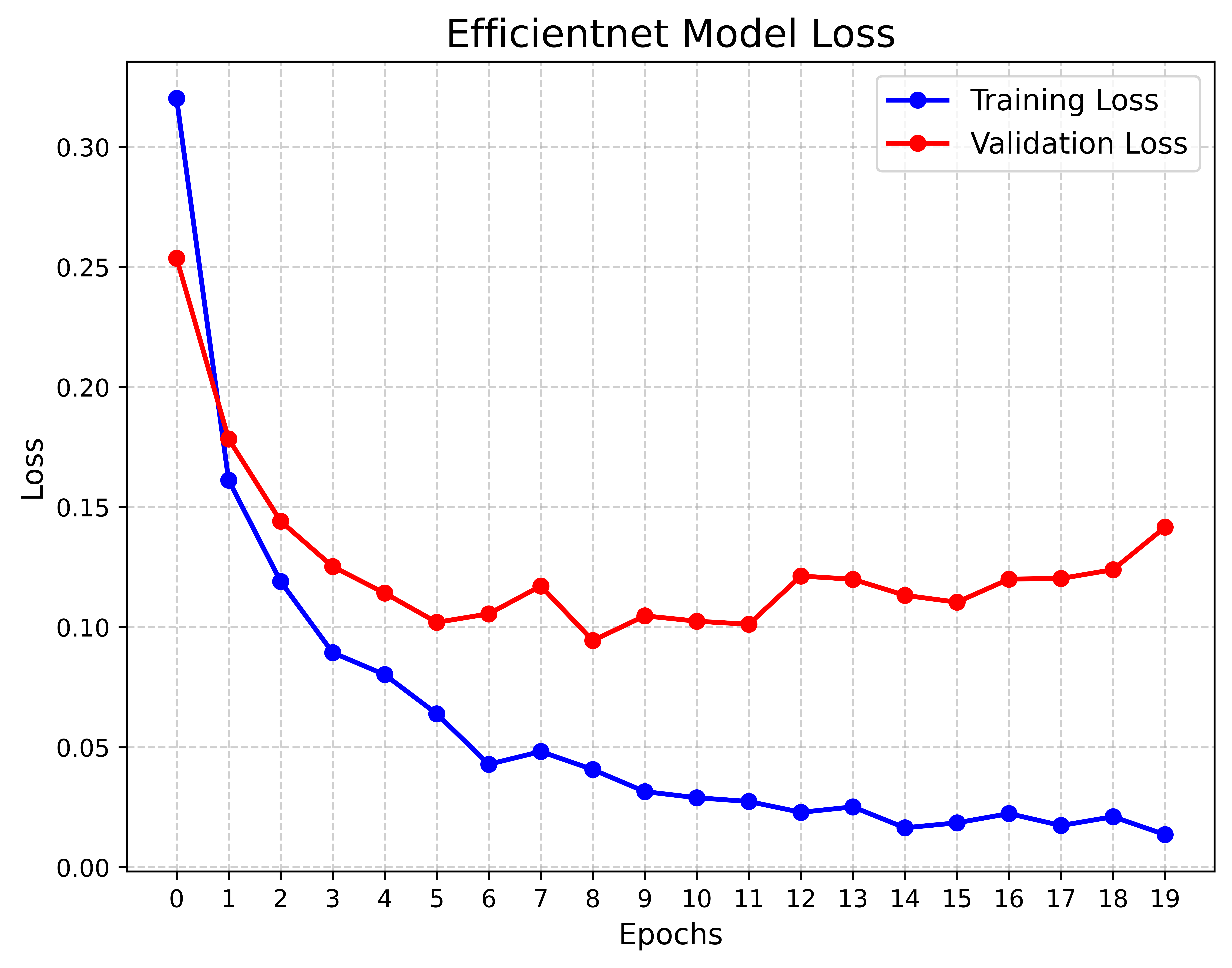
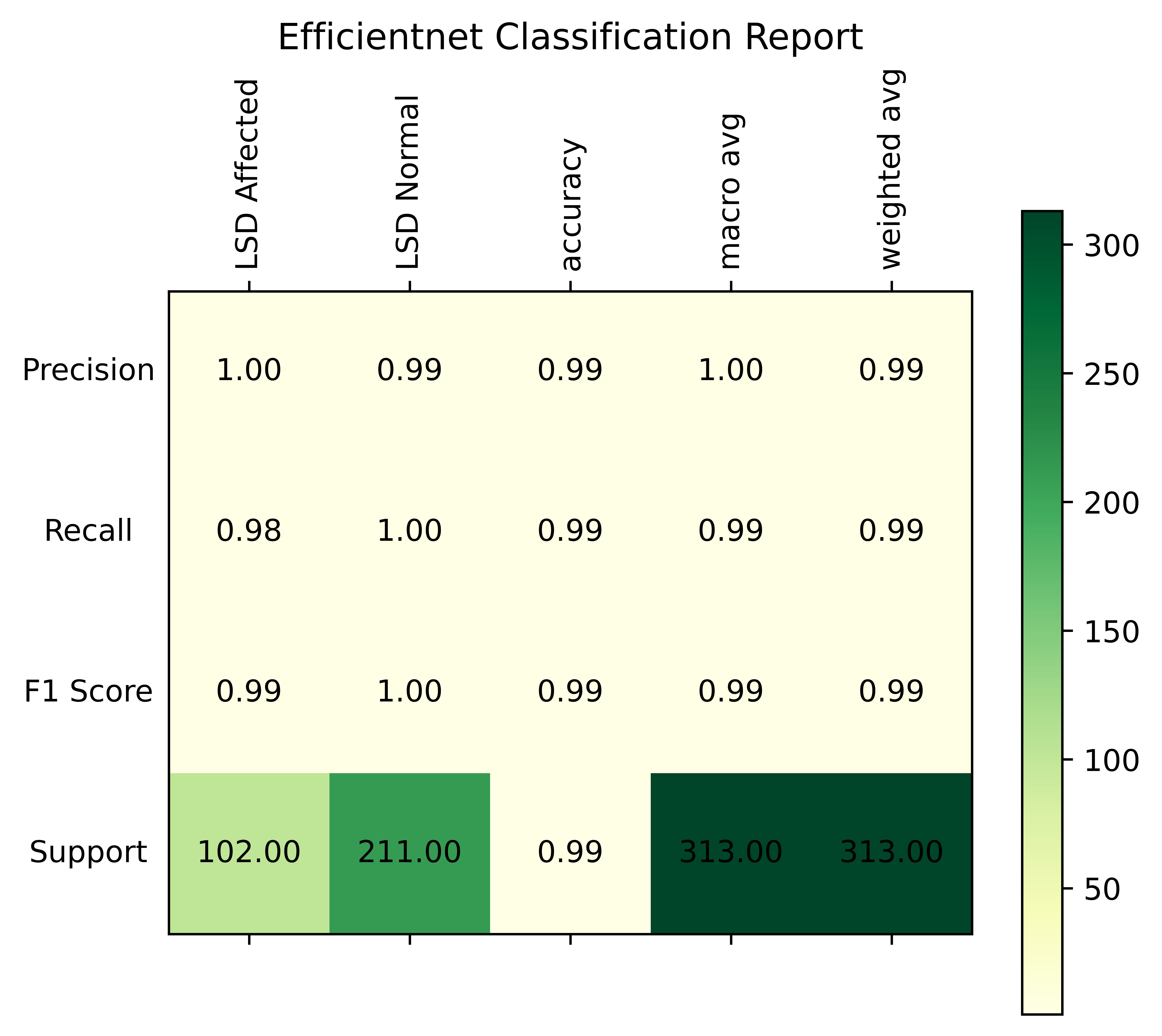
The proposed model is tested on a high-performance machine equipped with 64 GB RAM, an NVIDIA RTX 4090 GPU, and a 13th Gen Intel Core i9 processor running Windows 11. For reproducibility and collaborative experiments, Google Colab is also utilized, offering seamless access to widely-used libraries, support for both GPU and TPU acceleration, and smooth integration with Google Drive for efficient data storage and sharing. The model is tested using the Lumpy skin image dataset, which is publicly available [14]. We use Compute Unified Device Architecture (CUDA) to enhance the computational efficiency of the model. It also increases the speed of the model-training process. The improved performance may be attributed to DL’s ability to extract rich, hierarchical information from photos of cattle impacted by LSD. The results presented clearly demonstrate that our DL-based techniques perform better than traditional ML models for early LSD detection. The robustness of the suggested method in successfully detecting and categorizing LSD cases is implied by its high accuracy (99%) and exceptional AUC, F1-score, precision, and recall. While the recall for healthy cattle is 1.0, indicating the model’s ability to catch all non-inflamed instances without any false negatives, the recall for inflamed livestock is 0.98, indicating that the model successfully recognizes 0.98% of inflamed cases, but it misses some. The model’s overall best performance is highlighted by the F1 score, which strikes a compromise between precision and recall, being 1.0 for healthy cattle and 0.99 for inflamed farm animals.
Figure 7. LUMPNet Model lossFigure 8. LUMPNet Classification Report
IV. Conclusion
The proposed work on detecting LSD in cattle through image data and AI demonstrates a comprehensive and effective workflow that integrates a novel AWDR optimizer with two major ML techniques. This work utilizes YOLOv11 for cattle detection and EfficientNet for accurately identifying LSD symptoms, particularly skin nodules, which are early indicators of the disease. The results show that the LUMPNet model performs exceptionally well, achieving 99% accuracy, high precision, recall, and F1 scores, confirming its reliability in detecting infected cattle. The systematic approach from dataset preparation to evaluation proves to be transformative for early disease detection, offering significant benefits to livestock management by enabling timely intervention and improving animal health outcomes. This research paves the way for a robust AI-driven solution in veterinary disease management, ensuring more efficient and accurate detection of diseases like LSD in cattle. LUMPNet model introduces a hybrid deep learning framework that integrates AWDR optimizer and YOLOv11 with the SAM to achieve both object detection and fine-grained segmentation of LSD-affected cattle. Unlike standard YOLOv11, which detects objects using bounding boxes alone, this approach enhances precision by segmenting lesion areas after dectection, making it particularly effective for medical image analysis.
References
[1] C. Krampe, P. T. Ingenbleek, J. K. Niemi, and J. Serratosa, “Designing precision livestock farming system innovations: A farmer perspective,” Journal of Rural Studies, vol. 111, Art. no. 103397, 2024.
[2] E. S. Tuppurainen, S. Babiuk, and E. Klement, “Introduction to lumpy skin disease,” in Lumpy Skin Disease, 2018, pp. 1–2.
[3] K. Adamu, T. Abayneh, B. Getachew, H. Mohammed, G. Deresse, M. Zekarias, W. Chala, and E. Gelaye, “Lumpy skin disease virus isolation, experimental infection, and evaluation of disease development in a calf,” Scientific Reports, vol. 14, no. 1, Art. no. 20460, 2024.
[4] H. F. Gouda and F. D. Abdallah, “Comparative performance of bagging and boosting ensemble models for predicting lumpy skin disease with multiclass-imbalanced data,” Scientific Reports, vol. 15, no. 1, Art. no. 39373, 2025.
[5] N. Ujjwal, A. Singh, A. K. Jain, and R. G. Tiwari, “Exploiting machine learning for lumpy skin disease occurrence detection,” in Proceedings of the 2022 10th International Conference on Reliability, Infocom Technologies and Optimization (Trends and Future Directions) (ICRITO). IEEE, 2022, pp. 1–6.
[6] C. Senthilkumar, G. Vadivu, and S. Neethirajan, “Early detection of lumpy skin disease in cattle using deep learning—a comparative analysis of pretrained models,” Veterinary Sciences, vol. 11, no. 10, Art. no. 510, 2024.
[7] S. Muhammad Saqib, M. Iqbal, M. Tahar Ben Othman, T. Shahazad, Y. Yasin Ghadi, S. Al-Amro, and T. Mazhar, “Lumpy skin disease diagnosis in cattle: A deep learning approach optimized with RMSprop and MobileNetV2,” PLOS ONE, vol. 19, no. 8, Art. no. e0302862, 2024.
[8] A. Ullah, A. Haider, Y. Rahulamathavan, M. W. Khokhar, M. A. Rohaim, F. Alam, N. Iqbal, and M. Munir, “A vision transformer model-integrated mobile application for early and accurate detection of lumpy skin disease in cattle,” Scientific Reports, 2025.
[9] K. Al-Salihi et al., “Lumpy skin disease: Review of literature,” Mirror of Research in Veterinary Sciences and Animals, vol. 3, no. 3, pp. 6–23, 2014.
[10] K. Ananda and P. K. MJ, “Machine learning-based detection of lumpy skin disease in cattle using deep feature extraction techniques,” in Proceedings of the 2025 International Conference on Intelligent and Innovative Technologies in Computing, Electrical and Electronics (IITCEE). IEEE, 2025, pp. 1–6.
[11] R. Raj, S. Panda, N. Nitya, D. Patel, and D. Muduli, “Automated diagnosis of lumpy skin diseases based on deep learning feature fusion,” in Proceedings of the 2023 14th International Conference on Computing Communication and Networking Technologies (ICCCNT). IEEE, 2023, pp. 1–4.
[12] Z. Liang, S. Wang, K. Yao, S. Ren, P. Cheng, M. Qu, X. Ma, X. Gao, X. Yin, X. Wang et al., “Lumpy skin disease virus ORF127 protein suppresses type I interferon responses by inhibiting K63-linked ubiquitination of TANK-binding kinase 1,” The FASEB Journal, vol. 38, no. 3, Art. no. e23467, 2024.
[13] C. Morgan-Davies, G. Tesnière, J.-M. Gautier, G. Jørgensen, E. González-García, S. Patsios, E. Sossidou, T. W. Keady, B. McClearn, F. Kenyon et al., “Exploring the use of precision livestock farming for small ruminant welfare management,” Animal, vol. 18, Art. no. 101233, 2024.
[15] A. Kirillov, E. Mintun, N. Ravi, H. Mao, C. Rolland, L. Gustafson, T. Xiao, S. Whitehead, A. C. Berg, W.-Y. Lo et al., “Segment anything,” in Proceedings of the IEEE/CVF International Conference on Computer Vision (ICCV), 2023, pp. 4015–4026.
[16] L.-H. He, Y.-Z. Zhou, L. Liu, W. Cao, and J.-H. Ma, “Research on object detection and recognition in remote sensing images based on YOLOv11,” Scientific Reports, vol. 15, no. 1, Art. no. 14032, 2025.
[17] B. Koonce, “EfficientNet,” in Convolutional Neural Networks with Swift for TensorFlow: Image Recognition and Dataset Categorization. Apress, 2021, pp. 109–123.